TrueGrid® BioMechanical Gallery
Read about how to get surface data for biological geometry.
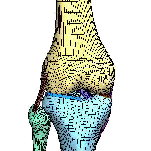
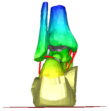
Mesh of Human Knee Ligaments
Dr. Jeff Weiss is an Associate Professor of Biomedical Engineering, at the University of Utah and the director of the Musculoskeletal Research Laboratories.
He has been modeling human soft tissue for many years. These images date from some of his mid-1990s finite element analyses of knee flexion, v-v rotation, tibial axial rotation, and a-p translation. Images provided courtesy of Jeff Weiss and his colleagues at the University of Utah. They retain all copyrights.
 |
 |
 |
 |
 |
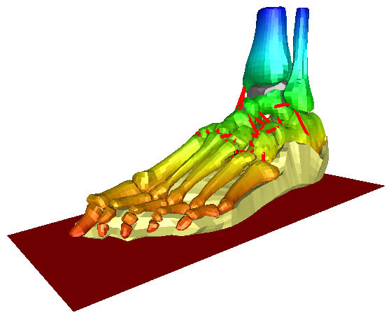
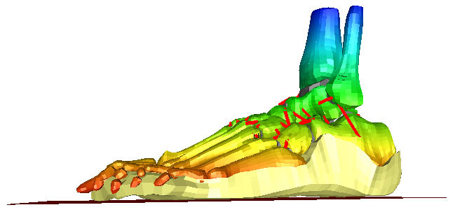
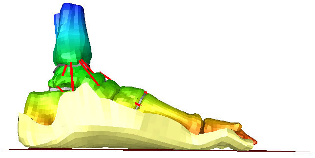
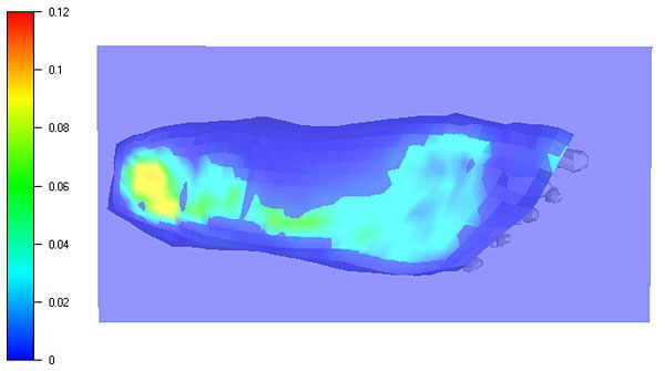
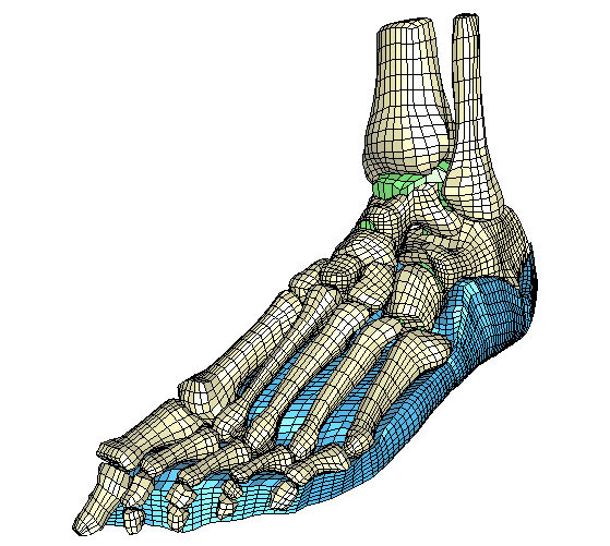
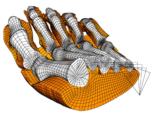
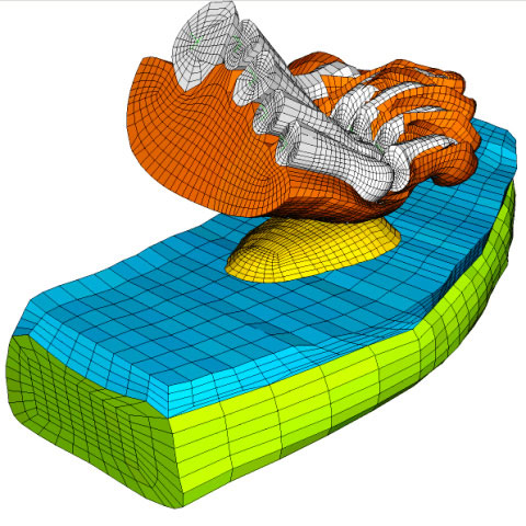
Finite Element Model of Human Foot
Dr. Bill Ledoux, Dr. Danny Camacho, and colleagues have created a finite element model of the human foot. The model includes bones, ligaments, plantar soft tissue, and cartilage. This work has been sponsored by VA Puget Sound and the University of Washington. Dr. Ledoux has graciously provided us with several views of his model and his results.
 |
 |
 |
 |
 |
 |
 |
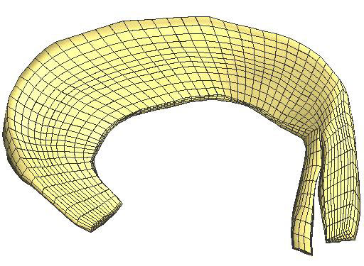
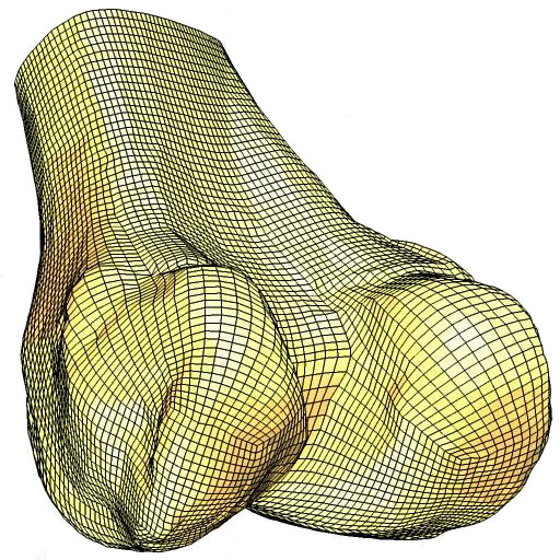
Finite Element Model of Human Femur
A single part in TrueGrid® is used to model a section of a human femur. The cross section of the mesh, best seen in the top view, reveals a double butterfly block structure. There are 190,560 nodes and 174,240 hexahedral (brick) elements in this model.
 |
 |
 |
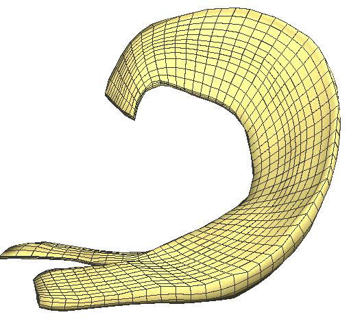
Mesh of the Human Heart
Over the years, more than one model of the human heart has been built using TrueGrid®. This is the earliest model, dating back to the mid-90s. The original data was obtained experimentally and then converted into polygonal surfaces before being read into TrueGrid®.
Researchers at Duke University have produced a model of portions of the human heart as they analyze the propagation of electrical current flow. Their results have been published in the on-line Journal of Circulation Research, sponsored by the American Heart Association.

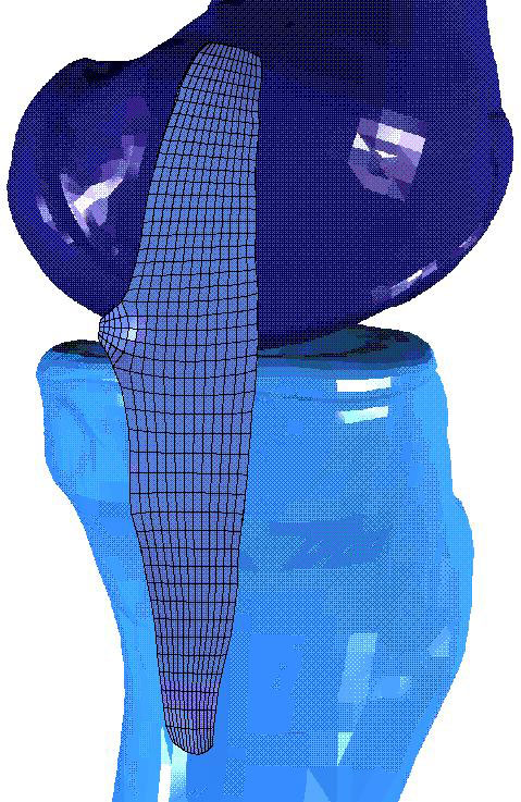
Mesh of Leg Bones and Implants
Dr. Donald Bartel is a Professor in the Department of Mechanical and Aerospace Engineering at Cornell University and Senior Scientist with the Laboratory for Biomedical Mechanics and Materials at the Hospital for Special Surgery in NYC. For those who wish to learn more about these organizations, their personnel and research, the Biomedical Mechanics program with the school of Mechanical and Aerospace Engineering Dept. at Cornell University has their own well organized web site.
For many years Dr. Bartel has studied the biomechanics and performance of bone-implant systems, focusing primarily on the hip and knee. These images are from current research on secondary fractures resulting from internal fixation of femoral neck fractures and the role of stem augmentation in constrained condylar knees.
A growing desire for statistically based analyses requires multiple physical models that emphasize some of TrueGrid®'s strengths. Its morphing and projection-based geometry creation, flexibility with surfaces and batch processing capabilities allow for efficient creation of comparable finite element meshes for different bones and implants. The images of the tibia show two implants for two bones created with essentially the same truegrid session file and minimal analyst input.
These images are provided courtesy of Dr. Bartel and his colleagues at Cornell University. They retain all copyrights.
 |
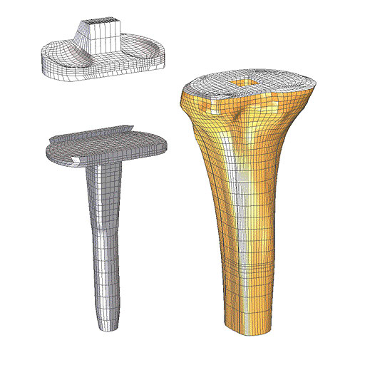 |
| Femur with Pins - secondary fractures resulting from internal fixation of femoral neck fractures | Tibia 1 with Implants - stem augmentation in constrained condylar knees |
 |
|
| Tibia 2 with Implants - stem augmentation in constrained condylar knees |
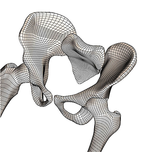
Mesh of Hip Bones
 |
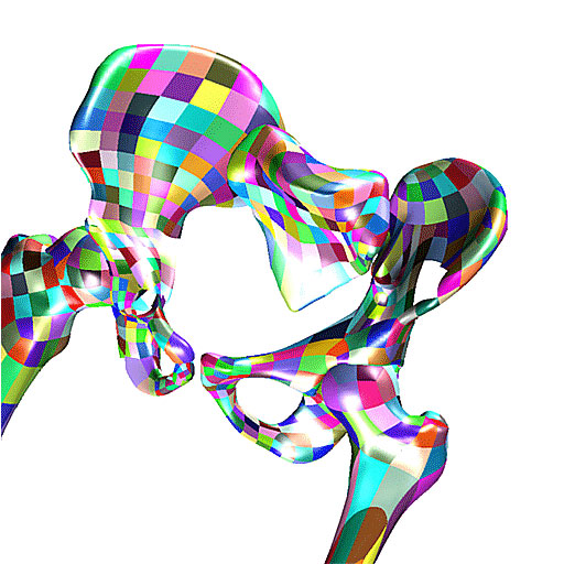 |
| Hex Mesh of Hip Bone | CAD Geometry of Hip Bone |
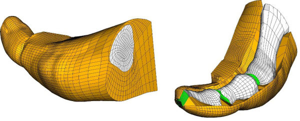
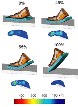
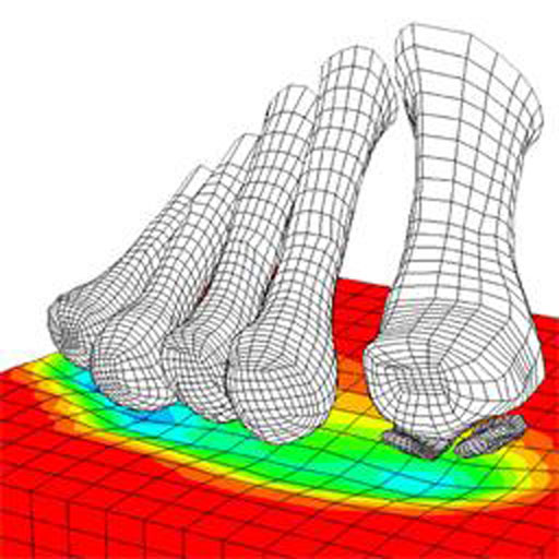
Model of Human Foot Walking
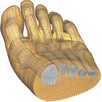
Sachin P. Budhabhatti has created a hexahedral mesh of a forefoot model for a current study directed by Dr. Peter R. Cavanagh at The Cleveland Clinic Foundation. These images are provided courtesy of Cavanagh Lab at Cleveland Clinic Foundation. They retain all the copyrights.
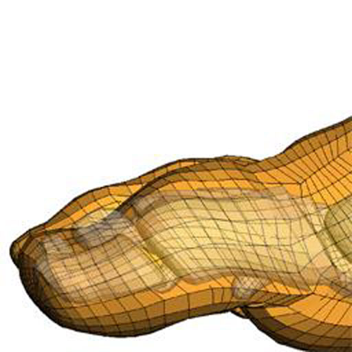
The first ray model was used to simulate various first ray deformities like hallux rigidus, arthrodesis, and footwear interventions. It includes 3 bones (metatarsal, proximal phalanx, and distal phalanx), soft-tissue, and cartilage. It was also used as a stepping stone to creating the forefoot mesh.
These images are provided courtesy of Cavanagh Lab at Cleveland Clinic Foundation. They retain all the copyrights. The study was directed by Dr. Peter R. Cavanagh at Cleveland Clinic. For more information about this mesh contact: Sachin P. Budhabhatti.
This model includes 17 bones (five metatarsals, five proximal phalanges, five distal phalanges, and two sesamoids) and soft tissue. The model is being used for morphing and footwear design purposes by Cavanagh lab.
 |
 |
 |
 |
| Foot Model Posterior View |
Foot Model Side View |
 |
 |
| Foot Model Bottom View |
Foot Model Results |
 |
 |
| Forefoot Model | Forefoot Model |
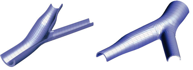
Mesh of an Arterial Bifurcation
This model was built by Sachin P. Budhabhatti for educational purposes.